

Welcome
Non-invasive and free from drugs and their side effects, low level laser therapy can be used on a wide group of patients.
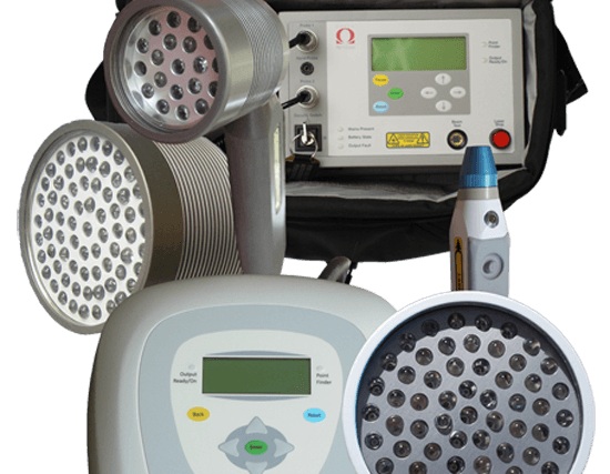
Omega Laser Systems have more than 25 years of research, developing and manufacturing experience in low level laser therapy.
